Figure 1. [The normal human retina fundus]. - Webvision - NCBI
Por um escritor misterioso
Last updated 20 setembro 2024
![Figure 1. [The normal human retina fundus]. - Webvision - NCBI](https://www.ncbi.nlm.nih.gov/books/NBK554706/bin/Archetecture_Fovea-Image006.jpg)
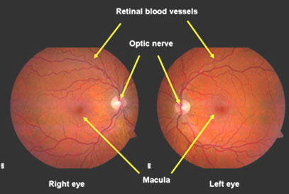
The normal human retina fundus photo shows the optic nerve (right), blood vessels and the position of the fovea (center).
![Figure 1. [The normal human retina fundus]. - Webvision - NCBI](https://www.pnas.org/cms/10.1073/pnas.2307380120/asset/9de33f2a-4bb0-4081-926d-bdb80222d13d/assets/images/large/pnas.2307380120fig01.jpg)
Cellular migration into a subretinal honeycomb-shaped prosthesis for high-resolution prosthetic vision
![Figure 1. [The normal human retina fundus]. - Webvision - NCBI](https://www.ncbi.nlm.nih.gov/books/NBK11556/bin/factsf2b.gif)
Facts and Figures Concerning the Human Retina - Webvision - NCBI Bookshelf
![Figure 1. [The normal human retina fundus]. - Webvision - NCBI](https://webvision.med.utah.edu/wp-content/uploads/2018/07/Fig-14-macula-lutea.jpg)
Simple Anatomy of the Retina by Helga Kolb – Webvision
![Figure 1. [The normal human retina fundus]. - Webvision - NCBI](http://webvision.med.utah.edu/imageswv/glaucretina.jpeg)
Simple Anatomy of the Retina : 네이버 블로그
![Figure 1. [The normal human retina fundus]. - Webvision - NCBI](https://media.springernature.com/m685/springer-static/image/art%3A10.1186%2Fs13024-023-00655-y/MediaObjects/13024_2023_655_Fig4_HTML.png)
Retinal ganglion cell repopulation for vision restoration in optic neuropathy: a roadmap from the RReSTORe Consortium, Molecular Neurodegeneration
![Figure 1. [The normal human retina fundus]. - Webvision - NCBI](https://www.frontiersin.org/files/Articles/687495/fphar-12-687495-HTML/image_m/fphar-12-687495-g003.jpg)
Frontiers Galectins in the Pathogenesis of Common Retinal Disease
![Figure 1. [The normal human retina fundus]. - Webvision - NCBI](https://media.springernature.com/full/springer-static/image/art%3A10.1038%2Fs41467-019-12917-9/MediaObjects/41467_2019_12917_Fig1_HTML.png)
Single-nuclei RNA-seq on human retinal tissue provides improved transcriptome profiling
![Figure 1. [The normal human retina fundus]. - Webvision - NCBI](https://www.mdpi.com/cells/cells-12-01987/article_deploy/html/images/cells-12-01987-g001.png)
Cells, Free Full-Text
![Figure 1. [The normal human retina fundus]. - Webvision - NCBI](https://www.ncbi.nlm.nih.gov/books/NBK11556/bin/factsf6.gif)
Facts and Figures Concerning the Human Retina - Webvision - NCBI Bookshelf
![Figure 1. [The normal human retina fundus]. - Webvision - NCBI](https://www.ncbi.nlm.nih.gov/books/NBK11553/bin/clinicalergf31b.jpg)
Figure 31b, [Optical coherence tomography (OCT) images]. - Webvision - NCBI Bookshelf
![Figure 1. [The normal human retina fundus]. - Webvision - NCBI](https://media.springernature.com/lw685/springer-static/image/art%3A10.1007%2Fs11042-022-13837-5/MediaObjects/11042_2022_13837_Fig1_HTML.png)
A survey on recent developments in diabetic retinopathy detection through integration of deep learning
Recomendado para você
-
Retina - American Academy of Ophthalmology20 setembro 2024
-
 About the Eye, Eye Care Atlanta, Retina Care Atlanta20 setembro 2024
 -
 Retinal Diseases: Types, Causes, Symptoms, Treatment, Outlook20 setembro 2024
 -
 Retina: 3 doenças hereditárias e raras. - CBCO20 setembro 2024
 -
 Normal Retina vs. Papilledema - Trial Exhibits Inc.20 setembro 2024
 -
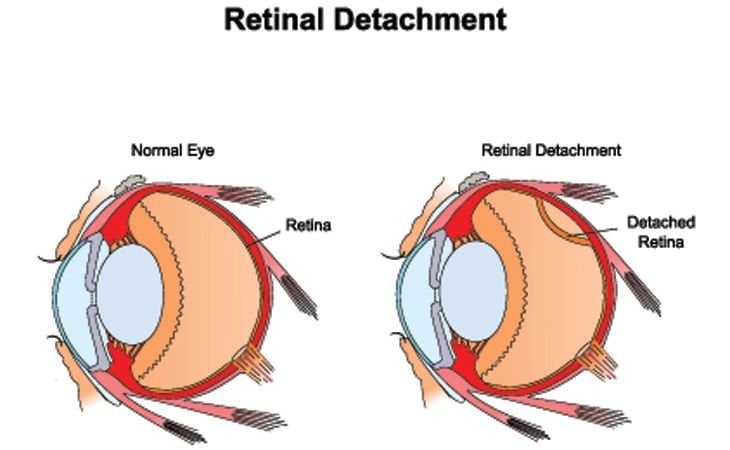
 Descolamento de Retina - Dra. Camila Otani20 setembro 2024
 -
 RETINA HOSPITAL20 setembro 2024
 -
 Detached Retina, Optometrist in Chicago, Illinois20 setembro 2024
 -
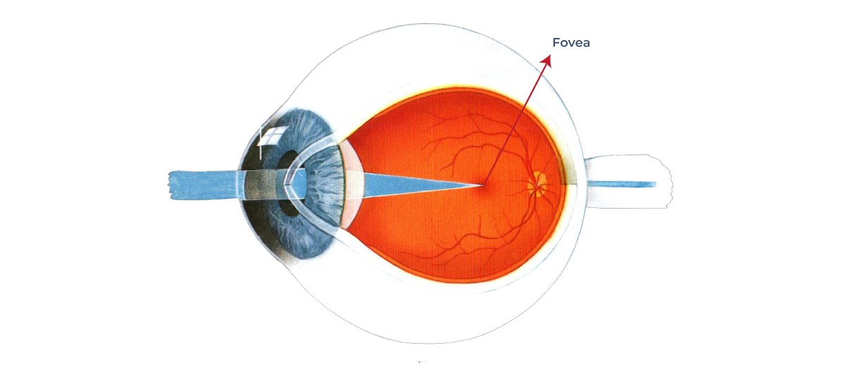 What is the fovea? – Front Range Retina20 setembro 2024
 -
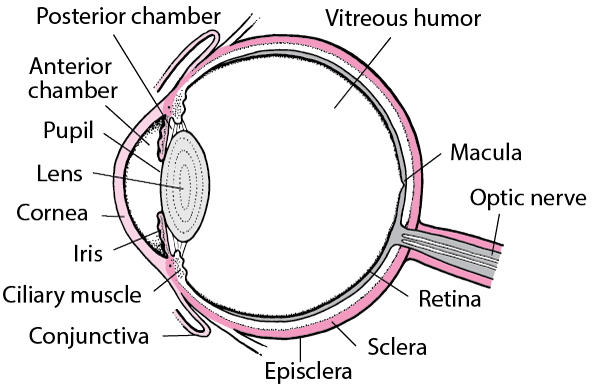 Structure and Function of the Eyes - Eye Disorders - Merck Manuals20 setembro 2024
você pode gostar
-
 Jaro From Subway Surfers coloring page20 setembro 2024
 -
 Rüzgarın kalbi dizisi 5. bölüm ne zaman yayınlanacak?20 setembro 2024
 -
 Download jogo xbox 360 gratis20 setembro 2024
 -
 How to Connect to The Fastest Server on Roblox! (Easy)20 setembro 2024
 -
 Harold Faltermeyer - Top Gun Anthem - RECORD SLEEVE ONLY (45RPM 7”) (RC275)20 setembro 2024
 -
 Withered Foxy — Minecraft head20 setembro 2024
 -
This goes crazy #skylandprodigy #scratchfnf #fnf #fnftest20 setembro 2024
-
Rotten Tomatoes - New Spider-People logos for Spider-Man: Across The # SpiderVerse - in theaters June 2.20 setembro 2024
-
 How the Five Eyes alliance fuels global surveillance20 setembro 2024
 -
Mobile Unit Summer of Joy Digital Program20 setembro 2024

